Is Laparoscopy the Gold Standard for the Diagnosis of Endometriosis?
By I.A. Brosens(a) and J.J. Brosens(b)
(a) Leuven Institute for Fertility and Embryology, Leuven, Belgium
(b) Department of Reproductive Sciences and Medicine, Division of Paediatrics, Obstetrics and Gynaecology, ICSM at Hammersmith Hospital, London, UK
* Corresponding author. Tel.: +32-16-270190; fax: +32-16-270197. E-mail address mailto:%20ivo.brosens@med.kuleuven.ac.be
For several decades laparoscopy has been the gold standard for the diagnosis of endometriosis. Indeed, the definition of endometriosis was created by the reflux concept of Sampson's (1) and consequently, visualization of hemorrhagic implants and, as final proof, biopsies showing glands and stroma have been the basis of the diagnosis. Today, there are major reasons to state that the definition and consequently the place of laparoscopy in the diagnosis should undergo a major revision.
First, the definition of endometriosis is based on the reflux concept and not on the original observation of specific endometrial activity at ectopic sites. Indeed, Sampson (2) discovered endometriosis by observing menstrual shedding in endometrial-like, tissue in ovarian chocolate cysts in two patients operated at the time of menstruation.
Secondly, the visual, mechanistic definition neglects to include the smooth muscle cell hyperplasia, which was accurately described by CuIlen (3) to occur along the Müllerian tract, primarily in the myometrium, the posterior fornix, the uterosacral ligaments and to some extent at other fibromuscular sites. It is now well recognized that smooth muscle differentiation is also a specific activity associated with basal endometrium. The present confusing terminology of deep, infiltrating and invasive endometriosis has been a consequence of restricting a disease process to a concept, which neglects the metaplastic changes of mesenchymal cells differentiating into smooth muscle cells (4). The so called deep, rectovaginal endometriosis in contrast with peritoneal and ovarian endometriosis does not correspond with the phenotype of superficial, but basal endometrium and presents all the morphological features of adenomyosis (5,6).
It is therefore logical to redefine endometriosis by the specific, functional changes associated with ectopic endometrial-like tissue. Regardless of the underlying etiology and pathogenesis, the phenotype of ectopic endometrial-like tissue is apparently determined by the surrounding microenvironment (7). Peritoneal and ovarian endometriosis have characteristics, albeit defective, of superficial endometrium and are functionally characterized by sex steroid hormone-dependent bleeding. in contrast, rectovaginal endometriosis, like diffuse uterine adenomyosis is similarly as basal endometrium characterized by inordinate smooth muscle differentiation and hyperplasia and nodule formation.
Moreover, there is increasing evidence that endometriosis is part of a pleiotropic reproductive disorder including aberrant eutopic endometrium, disruption of normal inner myometrial peristalsis, proinflammatory state of the peritoneum, abnormal ovarian steroidogenesis and impaired oocyte maturation (8). The normal sex steroid hormone response of the Müllerian tract is disrupted and the presence of endometriotic implants is only one aspect of the pleiotropic reproductive disorder. Consequently, if the visual concept of endometriosis is being replaced by a functional definition, the method of diagnosis should also undergo a major revision.
There are also clinical reasons to review the place of traditional laparoscopy in the diagnosis of endometriosis. Whilst not classified as major surgery, laparoscopy is an invasive and expensive procedure. It requires general anesthesia of the patient and full operating theatre facilities. The transabdominal approach is responsible for approximately 50% of the complications (9,10). Injury to a major blood vessel can be catastrophic with a reported mortality of 15% and the offending instrument is the Veress needle as often as the trocar (11). The diagnosis of endometriosis by laparoscopy has therefore never been practical. The delay in the diagnosis of endometriosis in patients with infertility and chronic pelvic pain Is 3.5 and 11.7 years respectively (12). The delay results in disease progression with increased risk of persistent disease and patient's anxiety and depression. Monitoring the evolution of the disease by laparoscopy has also not been routine practice. On the contrary, the role of purely diagnostic laparoscopy is being gradually eliminated from the contemporary management of endometriosis, in which suspected lesions are treated surgically when they are first seen. A recent Canadian study showed that this approach results in a modest increase in subsequent pregnancy rates (13). The results of the study remain controversial, but the most consistent finding in similar studies is that destroying visible implants fails to cure the disease.
Therefore, there are major reasons to revise the diagnostic approach of endometriosis. Alternatives to standard laparoscopy for the diagnosis of endometriosis have already been proposed or are in development. Recently, a new office procedure based on the transvaginal access and the use of saline as distension medium, called transvaginal hydrolaparoscopy (THL), has been proposed as a more suitable screening method for early diagnosis of endometriosis in patients with infertility or chronic pelvic pain (14). The safety factors include the use of local anesthesia. transvaginal access, needle technique and saline for distension. The systematic use of saline for distension makes THL a much more sensitive technique for the diagnosis of adhesions. For instance, examination of the ovaries by THL in patients with mild endometriosis revealed 50% more periovarian adhesions compared to standard laparoscopy (15). In addition, THL is likely to restore the normal decision making process of a surgical procedure which proceeds from diagnosis to evaluation of the treatment options with the patient and ultimately a planned surgical procedure. Conventional T2-weighed MR imaging, on the other hand, is an accurate technique to detect adenomyotic hyperplasia in the myometrium. posterior fornix and uterine ligaments (16). This imaging technique can also detect hemorrhagic lesions if greater than 4 mm (17). The development of ultrasensitive endocavitary uterine and rectal MR receiver coils is likely to facilitate detection of early lesions along the Müllerian tract. The technique is noninvasive, is more cost effective than laparoscopy, and can be repeated when indicated.
In conclusion, not only the definition, but also the diagnosis of endometriosis should undergo a major revision. Pelvic endoscopy remains a useful technique to visualize and document the superficial, hemorrhagic type of endometriosis and adhesions, but fails to reveal the adenomyostic, nodular type and, even more important, the pleiotropic reproductive abnormalities. If the visual concept of endometriosis is no longer tenable and endometriosis is apparently part of a pleiotropic reproductive disorder, characterized by disruption of normal sex steroid hormone dependent differentiation process in the Müllerian tract, it is more likely that in the near future a combination of techniques is desirable for the diagnosis of the disease endometriosis/adenomyosis.
REFERENCES
Sampson JA (1927) Peritoneal endometriosis due to the menstrual dissemination of endometrial tissue into the peritoneal cavity. Am J Obstet Gynecol 14: 422-69.
Sampson JA (1921) Perforating hemorrhagic (chocolate) cysts of the ovary. Arch Surg 1921: 245-323
Cullen TS (1920) The distribution of adenomyoma containing uterine mucosa. Arch Surg 1: 215-83.
Fujii S, Konishi I, Mon T (1989) Smooth muscle differentiation at endometrio-myometrial junction. An `ultrastructural study. Am J Obstet Gynecol 163: 105-12
Brosens IA (1994) New principles in the management of endometriosis. Acta Obstet Gynecol Scand Suppl 159:18-21.
Donnez J, Nisolle M, Smoes P. Gillet N, Beguin 5, Casanas-Roux F (1996) Peritoneal endometriosis and "endometriotic" nodules of the rectovaginal septum are two different entities. Ferti Steril 66: 362-8.
Tabibzadeh S. Sun XZ, Kong OF, Kasnic G, Miller J, Satyaswaroop PG (1993) Induction of a polarized micro-environment by human T cells and interferon-gamma in three dimensional spheroid cultures of human endometrial epithelial cells. Hum Reprod. 8: 182-92.
Brosens JJ, Brosens IA. From a visual to a functional diagnosis of endometriosis: implications for the diagnosis. Am J Obstet Gynecol (submitted for publication).
Chapron C, Querleu D, Bruhat M-A, Madelenat P, Fernandez H, Pierre F, Dubuisson J-B (1998) Surgical complications of diagnostic and operative gynaecological laparoscopy : a series of 29 966 cases. Hum Reprod 13: 867-72.
Jansen FW, Kapiteyn K, Trimbos-Kemper T, Hermans J, Trimbos JB (1997) Complications of laparoscopy: prospective multicentre observational study. Br J Obstet Gynaecol 104: 595-600.
Baadsgaard SE, Bille S, Egeblad K (1989) Major vascular injury during gynecologic laparoscopy. Acta Obstet Gynecol Scand 68: 283-5.
Dmowski WP, Lesniewicz R, Rana N, Pepping. Changing trends in the diagnosis of endometriosis: a comprehensive study of women with pelvic endometriosis presenting with chronic pelvic pain or infertility. Fertil Steril 67: 238-43.
Marcoux S, Maheux R, Bérubé S and the Canadian Collaborative Group on Endometriosis. (1997) Laparoscopic surgery in infertile women with minimal or mild endometriosis. N Eng J Med 337: 217-22.
Gordts S, Campos R, Rombauts L, Brosens I (1998) Transvaginal hydrolaparoscopy as an outpatient procedure for infertility investigation. Hum Reprod 13: 99-103.
Campo R, Gordts S, Rombauts L, Borsens I (1999) Diagnostic accuracy of transvaginal hydrolaparoscopy in infertility. Fertil Steril 71: 1157-60.
Mark AS, Hricak H, Heinrichs LW, Hendrickson MR, Winkler ML, Bachica JA, Stickler JE (1987) Adenomyosis and leiomyoma: differential diagnosis with MR imaging. Am J Radiology 163: 527-9
Takahashi K, Okada s, Oszaki T, Kitao M, Sugimuri K (1994) Diagnosis of pelvic endometriosis by magnetic resonance imaging using "fat-saturation" technique. Fertil Steril 62: 973-7.
ISGE News
Editor: James E. Carter, M.D., Ph.D., F.A.C.O.G
ISGE Secretariat:
Spaarne Hospital
P.O. Box 1644
2003 BR Haarlem
The Netherlands
check out ISGE News online at
http://www.obgyn.net/medical.asp
© www.EndometriosisZone.org
BLOG DOKTER SPESIALIS KEBIDANAN DAN PENYAKIT KANDUNGAN ( Obstetric's & Gynecologist Blog ) Sumatera Barat.,Indonesia
SAVE YOUR BABY'S, SAVE NEXT GENERATION'S
SAVE YOUR BABY'S, SAVE NEXT GENERATION'S
Search This Blog
- Universitas Andalas Website
- TRIGEMINAL NEURALGIA LECTURES AND TREATMENT
- Maternal and Child :Research and Article
- dr Firman. Abdullah SpOG/ OBGYN .Personal Edition
- dr Firman Abdullah SpOG / ObGyn.com
- Dr Djohanas Djohan Abdullah Memorial Hospital.com
- Bukittinggi International Hospital.com
- Aliansi Rakyat Anti Korupsi Bukitinggi.com
Jam Gadang.Bukittinggi. Sumatera Barat .Indonesia
24.jpg)

Bung Hatta statue ,Bukittinggi
About me.....
IKATAN DOKTER INDONESIA (IDI).Sumatera Barat

INDONESIAN MEDICAL ASSOCIATION
ASSALAMUALAIKUM........
dr Firman Abdullah SpOG / OBGYN
Peer - Review..Cyberounds
Blog Archive
-
►
2008
(1)
- ► March 2008 (1)
-
▼
2009
(387)
- ► April 2009 (87)
-
▼
May 2009
(91)
- World Health Day 2009 ' Save Lives. Make hospitals...
- Pelvic exercises 'can help with childbirth and rec...
- More hysterectomy patients 'keeping cervix'
- Ovary removal 'may increase associated health risks'
- 'Fat gene' associated with PCOS
- Obese women 'at increased risk of birth defects'
- Resolution on Female Genital Mutilation
- Newborn babies have got rhythm, according to study
- Breastfeeding 'key during first six months'
- Mothers 'need breastfeeding support'
- Baby dies after receiving kiss infection
- Yoga 'can reduce menopause symptoms'
- ACOG President Advises Against Unnecessary Obstacl...
- Asoprisnil looks promising for endometriosis and u...
- Drospirenone and estradiol: a new option for the p...
- Endometriosis Surgery, State of the Art
- A call for centres of excellence to treat endometr...
- A Gonadotrophin-releasing Hormone Agonist compared...
- Accuracy of laparoscopic diagnosis of endometriosi...
- ACOG issues new practice bulletin on chronic pelvi...
- Adhesions in relation to laparoscopic surgery for ...
- Adolescent endometriosis
- Aromatase in endometriosis
- The Endometriosis Coping Zone Bowel Symptoms
- Changes in Immune and Endocrine System in Women wi...
- EndometriosisZONE.org Current Concepts and Researc...
- Dietary modification to alleviate endometriosis sy...
- Endometriosis does not impair obstetric outcome
- Endometriosis of the rectovaginal septum
- Endometriosis: The Four Pillars of Healing
- Endometriosis: the importance of early diagnosis a...
- GnRH Analogues in the Management of Endometriosis
- Is Laparoscopy the Gold Standard for the Diagnosis...
- Laparoscopic intraperitoneal injection of human in...
- Laparoscopic surgery helps relieve endometriosis pain
- Laterality of Endometriosis
- Link Between Migraine, Endometriosis Found
- Many women in Germany prefer long-cycle oral contr...
- Patterns of Understanding the Genetics of Endometr...
- Pre and post operative medical therapy for endomet...
- EndometriosisZONE.org Progesterone resistence in e...
- Radical endometriosis surgery is 'effective'
- Surgical Treatment For Endometriosis: Dr. Togas Tu...
- Tips for Dealing with Hot Flashes and Night Sweats...
- The LUNA procedure has no effect on endometriosis ...
- The Problem with Adhesions
- Extraperitoneal endometriosis, catamenial pneumoth...
- Managing endometriosis in teenagers
- Probable neuroimmunological link between Toxoplasm...
- What Causes Schizophrenia?
- CMV - Cytomegalovirus
- How the Herpes Simplex Virus Works
- Minilaparotomy and endoscopic techniques for tubal...
- Oral contraceptives for functional ovarian cysts
- Oral contraceptive pill as treatment for primary d...
- Steroidal contraceptives: effect on bone fractures...
- Prenatal administration of progesterone for preven...
- Minilaparotomy and endoscopic techniques for tubal...
- Danazol for pelvic pain associated with endometriosis
- Danazol for heavy menstrual bleeding
- Immersion in water in labour and birth
- Phenobarbital prior to preterm birth for preventin...
- Hysterectomy versus hysterectomy plus oophorectomy...
- Early postnatal discharge from hospital for health...
- Dehydroepiandrosterone (DHEA) supplementation for ...
- Total versus subtotal hysterectomy for benign gyna...
- Antibiotics for prelabour rupture of membranes at ...
- Abdominal surgical incisions for caesarean section
- Caesareans associated with fewer subsequent pregna...
- RCOG releases updated guidance on air travel durin...
- Too much exercise in early pregnancy may cause pre...
- Use of steroid in preterm birth appears safe
- Saving women's lives
- Vitamin E appears to relieve painful periods and r...
- Planned Caesarean decreases risk of complications ...
- Communicating the health risks and benefits of rep...
- RCOG Response to LIFE Claims on Link Between Abort...
- Elevated Rising CA 125 with Adenomyosis ---------...
- Ovarian Endometrioma Associated With Extremely Ele...
- Ovarian cancer in a woman previously diagnosed wit...
- Elevation of tumour marker CA-125 in serum & body ...
- MULTI-FOCAL EXTRA-UTERINE ENDOMETRIAL STROMAL SARC...
- LAPAROSCOPY INFORMATION
- Screening for vaginal shedding of cytomegalovirus ...
- Epstein–Barr Virus and Cytomegalovirus in Autoimmu...
- Systemic lupus erythematosus in adults is associat...
- A FOCUS ON FIBROIDS:
- Herpesvirus, cytomegalovirus, human sperm and assi...
- Influence Of The Menstrual Cycle On The Female Brain
- Cervical cancers after human papillomavirus vaccin...
- Health and Nutritional Benefits from Coconut Oil: ...
- ► August 2009 (54)
- ► September 2009 (21)
- ► November 2009 (4)
- ► December 2009 (11)
-
►
2010
(45)
- ► January 2010 (6)
- ► February 2010 (11)
- ► March 2010 (1)
- ► April 2010 (7)
- ► November 2010 (2)
-
►
2011
(4)
- ► February 2011 (2)
- ► March 2011 (2)
FEEDJIT Live Traffic Feed
Discussion Board
FEEDJIT Live Traffic Map
FEEDJIT Recommended Reading
FEEDJIT Live Page Popularity
dr Firman Abdullah SpOG / OBGYN

Subscribe to:
Post Comments (Atom)
BMI CALCULATOR
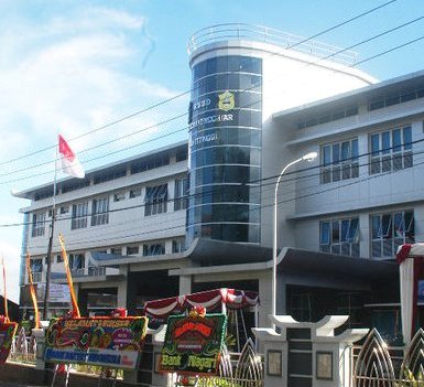
ACHMAD MOCHTAR GENERAL HOSPITAL BUKITTINGGI

RUMAH SAKIT ACHMAD MOCHTAR BUKITTINGGI
Firman Abdullah Bung
drFirman Abdullah SpOG / ObGyn

KELUARGA BESAR TNI-AD
Dr Firman Abdullah SpOG/ OBGYN, Bukittinggi, Sumatera Barat ,Indonesia
Bukittinggi , Sumatera Barat , Indonesia

Balaikota Bukittinggi
dr Firman Abdullah SpOG / OBGYN

Ngarai Sianok ,Bukittinggi, Sumatera Barat.Indonesia

Brevet in Specialist Obstetric's & Gynecologist 1998

dr Firman Abdullah SpOG/ObGyn


Dokter Spesialis Kebidanan dan Penyakit Kandungan . ( Obstetric's and Gynaecologist ) . Jl.Bahder Johan no.227,Depan pasar pagi ,Tembok .Bukittinggi 26124 ,HP:0812 660 1614. West Sumatra,Indonesia
Sikuai Beach ,West Sumatra ,Indonesia


Fort de Kock, Bukittinggi


No comments:
Post a Comment